
Monday, May 11, 2020
Case of the Week 590
This week's fascinating case was donated by Dr. Graham Hickling. He captured the following scene that he entitled "Hatching". What are these newborns?
 See them here LIVE (fast motion):


Sunday, May 10, 2020
Answer to Case 590
Answer to Parasite Case of the Week 590: Ixodes scapularis larvae, freshly hatched from an egg mass laid by a captive female. This fascinating video was generously donated by Dr. Graham Hickling who does research on hard tick biology.
While there isn't enough information present to specifically identify these ticks, you can say a few things about them from the video:
We've actually seen these little wonders before in a different blog post:
HERE is momma tick laying her eggs, with a video showing the action HERE. I HIGHLY recommend this video - it is fascinating! You may want to check out the other great videos on this site: https://vimeo.com/user1369895. Thanks again to Graham for sharing his 'babies' and passion for acarology with the rest of the world.
While I. scapularis tick larvae won't usually hatch until this summer, the nymphs that have over-wintered from last year are now out, so be sure to take precautions against tick bites when outdoors!
While there isn't enough information present to specifically identify these ticks, you can say a few things about them from the video:
- The have only 6 legs, and are therefore larvae.
- In North America, the long mouth parts and ovoid body would be most consistent with Ixodes species.
We've actually seen these little wonders before in a different blog post:
HERE is momma tick laying her eggs, with a video showing the action HERE. I HIGHLY recommend this video - it is fascinating! You may want to check out the other great videos on this site: https://vimeo.com/user1369895. Thanks again to Graham for sharing his 'babies' and passion for acarology with the rest of the world.
While I. scapularis tick larvae won't usually hatch until this summer, the nymphs that have over-wintered from last year are now out, so be sure to take precautions against tick bites when outdoors!
Monday, May 4, 2020
Case of the Week 589
Happy Monday everyone! It's our first Monday of the Month, and time for our monthly case from Idzi Potters and the Institute of Tropical Medicine, Antwerp.
The following objects were seen in an iron hematoxylin-stained stool preparation from an asymptomatic patient with frequent travels to Asia. The objects measure 10-20 micrometers in greatest dimension.


Identification?
The following objects were seen in an iron hematoxylin-stained stool preparation from an asymptomatic patient with frequent travels to Asia. The objects measure 10-20 micrometers in greatest dimension.


Identification?
Sunday, May 3, 2020
Answer to Case 589
Answer to Parasite Case of the Week 589: Iodamoeba buetschlii cysts and trophozoites.
Note that the trophozoites and cysts have a single nucleus with a large, often eccentric, karyosome. Cysts characteristically have a large glycogen-filled cytoplasmic vacuole that is highlighted when using iodine on a wet mount preparation. The glycogen vacuole does not stain with trichrome or iron hematoxylin, and instead appears as an empty space as in this case:

This organism gets its genus name from its iodine staining property (iod + amoeba), whereas the species name honors the German protistologist, Johann Adam Otto Bütschli. Note that the term "buetschlii" is used when citing a formal scientific name rather than "bütschlii" as per nomenclature standards.
Blaine pointed out there there were also some Blastocystis along for the ride (or vice versa):
 Old One also noted that the large vacuole looks a bit like a mask, which is a good reminder to wear a mask when going out in public to protect others around you.
Note that the trophozoites and cysts have a single nucleus with a large, often eccentric, karyosome. Cysts characteristically have a large glycogen-filled cytoplasmic vacuole that is highlighted when using iodine on a wet mount preparation. The glycogen vacuole does not stain with trichrome or iron hematoxylin, and instead appears as an empty space as in this case:

This organism gets its genus name from its iodine staining property (iod + amoeba), whereas the species name honors the German protistologist, Johann Adam Otto Bütschli. Note that the term "buetschlii" is used when citing a formal scientific name rather than "bütschlii" as per nomenclature standards.
Blaine pointed out there there were also some Blastocystis along for the ride (or vice versa):

Monday, April 20, 2020
Case of the Week 588
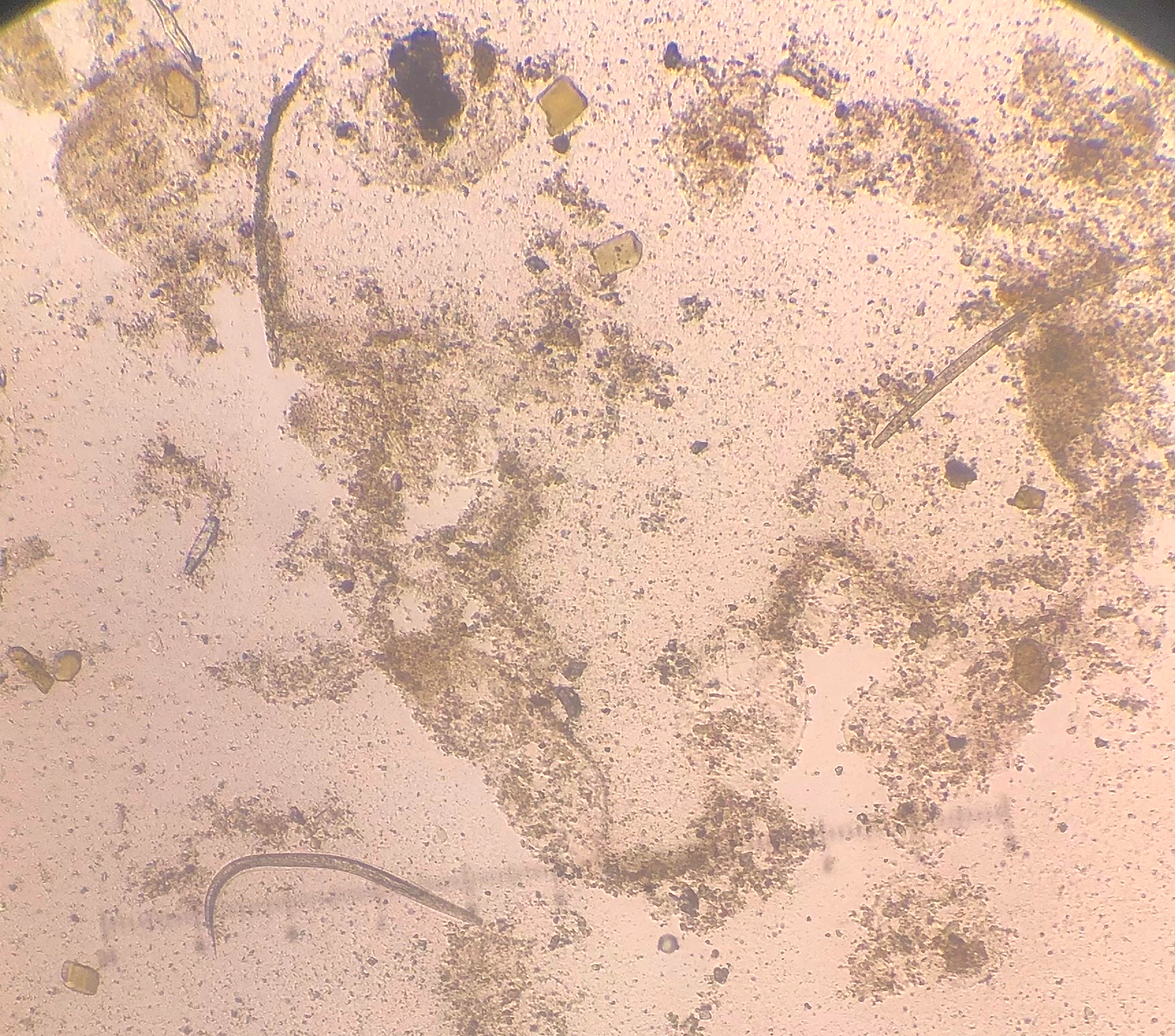
This week's case was donated by the very astute microbiology laboratory at the MetroHealth System who detected an important - but unexpected - finding. The patient is an elderly man on inhaled bronchodilators and steroids for persistent eosinophilic asthma who presented with fever, dry cough and shortness of breath. Reverse transcription PCR (RT-PCR) tests for circulating respiratory viruses were negative. However, blood cultures grew a number of bacteria including Gram negative bacilli, and the following were noted in a stool ova and parasite exam:


An additional finding was seen on the sheep blood agar plate of the sputum culture:

Review of the sputum showed the following:


What parasite is present here?


An additional finding was seen on the sheep blood agar plate of the sputum culture:

Review of the sputum showed the following:


What parasite is present here?
Sunday, April 19, 2020
Answer to Case 588
Answer to Parasite Case of the Week: Strongyloides stercoralis
This is a great teaching case from both the clinical and microbiology standpoint. As many of you pointed out, the clinical presentation is classic for Strongyloides hyperinfection with eosinophilic "asthma" (likely Loeffler's syndrome due to filariform (L3) larvae leaving the gut and migrating to the lung), receipt of an immunosuppressive agent (which decreases the host's ability to control the infection), and recurrent Gram negative bacteremia. As Harsha mentioned, the "Gram negative bacteria 'piggy back' on the larvae as they autoinfect entering from gut into blood setting up another cycle." This is why patients with hyperinfection syndrome commonly present with recurrent bacteremia and/or meningitis.
The microbiology findings are also classic. In this impressive case, we see many rhabditiform (L1) larvae in the stool. Filariform (L3) larvae may also be seen, although they are not highlighted in this case. The rhabditiform (L1) larvae of S. stercoralis have a short buccal canal (think S for strongy, and S for short), which allows them to be differentiated from the L1 larvae of the hookworms (which have a long buccal canal). Hookworm L1 larvae can occasionally be seen in stool specimens when the specimen is not collected into a fixative and is allowed to sit for some time before examination. Therefore, being able to differentiate between hookworm and S. stercoralis L1 larvae can be important. In this case, you can nicely see the short buccal canal, thus confirming this as S. stercoralis:

We also have the respiratory findings which support the diagnosis of strongyloidiasis, given that hookworm infection does not produce ongoing respiratory symptoms. Seen here are S. stercoralis L3 larvae, and also eggs! Presumably the eggs came from adults in the lung, which must have matured from the L3 larvae. Very cool. The bacterial trail from a migrating larva on the blood agar plate is another classic microbiology finding.
On a final note, I'd like to give credit to the astute internal medicine resident, Dr. Guoyou Chen, who thought to order the stool parasite exam based on the clinical findings of eosinophilic "asthma" and Gram negative bacteremia in the setting of immune compromise. Thanks also to Lorraine Sykes and the Microbiology laboratory for contributing this fascinating case!
This is a great teaching case from both the clinical and microbiology standpoint. As many of you pointed out, the clinical presentation is classic for Strongyloides hyperinfection with eosinophilic "asthma" (likely Loeffler's syndrome due to filariform (L3) larvae leaving the gut and migrating to the lung), receipt of an immunosuppressive agent (which decreases the host's ability to control the infection), and recurrent Gram negative bacteremia. As Harsha mentioned, the "Gram negative bacteria 'piggy back' on the larvae as they autoinfect entering from gut into blood setting up another cycle." This is why patients with hyperinfection syndrome commonly present with recurrent bacteremia and/or meningitis.
The microbiology findings are also classic. In this impressive case, we see many rhabditiform (L1) larvae in the stool. Filariform (L3) larvae may also be seen, although they are not highlighted in this case. The rhabditiform (L1) larvae of S. stercoralis have a short buccal canal (think S for strongy, and S for short), which allows them to be differentiated from the L1 larvae of the hookworms (which have a long buccal canal). Hookworm L1 larvae can occasionally be seen in stool specimens when the specimen is not collected into a fixative and is allowed to sit for some time before examination. Therefore, being able to differentiate between hookworm and S. stercoralis L1 larvae can be important. In this case, you can nicely see the short buccal canal, thus confirming this as S. stercoralis:

We also have the respiratory findings which support the diagnosis of strongyloidiasis, given that hookworm infection does not produce ongoing respiratory symptoms. Seen here are S. stercoralis L3 larvae, and also eggs! Presumably the eggs came from adults in the lung, which must have matured from the L3 larvae. Very cool. The bacterial trail from a migrating larva on the blood agar plate is another classic microbiology finding.
On a final note, I'd like to give credit to the astute internal medicine resident, Dr. Guoyou Chen, who thought to order the stool parasite exam based on the clinical findings of eosinophilic "asthma" and Gram negative bacteremia in the setting of immune compromise. Thanks also to Lorraine Sykes and the Microbiology laboratory for contributing this fascinating case!
Monday, April 13, 2020
Case of the Week 587
This week's case features objects seen in a concentrate from a diarrheic stool specimen. The objects measure approximately 60 micrometers in greatest dimension. Identification?


Sunday, April 12, 2020
Answer to Case 587
Answer to Parasite Case of the Week 587: Balantioides (formerly Balantidium, Neobalantidium) coli. Note the characteristic circumferential cilia and large "kidney bean" shaped macronucleus:
 I was impressed by how many people knew about the complicated taxonomy of this organism. Blaine Mathison and I had published on Neobalantidium coli in our Medical Parasitology Taxonomy Update (J Clin Microbiol. 2019;57:e01067-18) only to discover shortly afterwards that Balantioides has priority over Neobalantidium (we are now working on a correction). From our research, we discovered that Alexeieff was the first to describe Balantioides to accommodate B. coli in 1931. Unfortunately this French manuscript went largely unnoticed for decades, and Pomajbikova et al. alternatively described Neobalantidium in 2013 to accommodate B. coli. We can credit Pomajbikova and Stensvold for later uncovering the 1931 manuscript for us and shedding light on current state of taxonomy of Balantioides coli.
I have a pdf of the 1931 French manuscript (with my own translation) if anyone is interested in it. I should note that my French is very rusty, so there may be some errors! Just send me a message at b_pritt@yahoo.com if you would like a copy.
I'd also encourage you to read all of the interesting comments from this case. There are a lot of great pearls in there.

I have a pdf of the 1931 French manuscript (with my own translation) if anyone is interested in it. I should note that my French is very rusty, so there may be some errors! Just send me a message at b_pritt@yahoo.com if you would like a copy.
I'd also encourage you to read all of the interesting comments from this case. There are a lot of great pearls in there.
Monday, April 6, 2020
Case of the Week 586
It's now time for our monthly case by Idzi Potters and the Institute of Tropical Medicine, Antwerp:
The following structures were found in an unstained wet mount from a Belgian patient without any recent travel abroad. The patient reports intestinal discomfort for approximately 1 week. The structures measure approximately 20 micrometers in length.


Identification?
The following structures were found in an unstained wet mount from a Belgian patient without any recent travel abroad. The patient reports intestinal discomfort for approximately 1 week. The structures measure approximately 20 micrometers in length.


Identification?
Sunday, April 5, 2020
Answer to Case 586
Answer to Parasite Case of the Week 586: Mushroom spores. Not a parasite.
As many of you pointed out, mushroom spores are a challenging parasite mimic, resembling hookworm eggs, Cystoisospora belli oocysts, and Giardia cysts. Fortunately, you can rule out the first two based on the small small size of these objects (20 micrometers long), whereas G. duodenalis can be ruled out by the lack of internal structures (e.g., nuclei, axoneme, median bodies). Two other viewers initially thought there was a lateral spine present (indicating Schistosoma mansoni) but later realized it was just an artifact.
 Florida Fan pointed out that I had previously shown a case of mushroom (morel) spores in May of 2019 (Parasite Case of the Week 544). Great memory Florida Fan! That case showed mushroom spores within an ascus, which was a fun twist.
Florida Fan also mentioned that Spring is here at our door, and now is the time to go out and hunt for mushrooms - something that can be done while social distancing! I hope you and your families are all staying safe (and sane) during this challenging time.
As many of you pointed out, mushroom spores are a challenging parasite mimic, resembling hookworm eggs, Cystoisospora belli oocysts, and Giardia cysts. Fortunately, you can rule out the first two based on the small small size of these objects (20 micrometers long), whereas G. duodenalis can be ruled out by the lack of internal structures (e.g., nuclei, axoneme, median bodies). Two other viewers initially thought there was a lateral spine present (indicating Schistosoma mansoni) but later realized it was just an artifact.

Florida Fan also mentioned that Spring is here at our door, and now is the time to go out and hunt for mushrooms - something that can be done while social distancing! I hope you and your families are all staying safe (and sane) during this challenging time.
Subscribe to:
Posts (Atom)


