This week's case was generously donated by Dr. Marc Couturier and Blaine Mathison. The patient is a young adult male from the South Central United States. No travel history is available. Review of his peripheral blood film reviewed the following:
Identification? What are the structures seen in the last 2 images?
Monday, June 15, 2020
Sunday, June 14, 2020
Answer to Case 595
Answer to Parasite Case of the Week 595: Plasmodium vivax with exflagellated microgametocytes. Free microgametes are seen.
The following excellent description was written by Marc Couturier and Blaine Mathison, the contributors of this case.
There are several features of this slide that are supportive for P. vivax.
First, many of the infected cells are reticulocytes (which are larger than mature RBCs), which P. vivax and P. ovale have predilection to infect. Second, the gametocytes are enlarged and fill almost the entire space within the parasitized RBC. Some of the infected RBCs also take the shape of neighbouring RBCs (distorted, pleomorphic); particularly in the areas where cells are dense. Third, the mature schizonts contained upwards of 18-20 merozoites with yellowish brown pigment present, which strongly points toward P. vivax, since the range of P. ovale tends to reach 14 merozoites at maximum. Fourth, some developing trophozoites were grossly amoeboid. Plasmodium ovale usually demonstrates some amoeboid characteristics, but it is usually more exaggerated with P. vivax. Notice also none of the infected RBCs were fimbriated (which, when present supports P. ovale but the absence of which does not rule-out P. ovale).
In the images provided, there was an absence of Schüffner’s stippling. There were some parasitized cells that did display stippling (not shown), but the majority did not. It is important to remember that stippling can be helpful to support an identification, but the absence of stippling does not rule-out the species that usually present with stippling (P. vivax and P. ovale). Stippling is a pH dependent feature and Giemsa stained slides are the best to display this due to the optimized pH (Wright and Wright-Giemsa are suboptimal for this).
Overall the RBC morphology was quite good, suggesting that the blood was processed soon after collection. However, we suspected that there was a delay in slide processing which likely led to exflagellation of microgametocytes, with free microgametes seen among the RBCs:
Exflagellation of microgametocytes in blood specimens post-collection can occur due to multiple factors including decreased temperature, decreased CO2, increased O2 and subsequent increased pH. Free microgametes can be confused with other blood parasites such as Trypanosoma or relapsing fever Borrelia spp. (RFB). The key differences between these organisms are as follows: RFB are thin, faint staining, and in thin films will typically maintain a sinusoidal amplitude. The staining is uniform throughout the length of the bacteria and the width of the spirochetes is also consistent along the length of the organism. As can be seen with the free microgametes, there is irregular amplitude, and chromatin visible within the structure (causing heterogenous thickness). Trypanosoma spp. on the other hand are irregularly shaped compared to RFB; however, they should be identified based on the presence of a characteristic large nucleus in the central part of the organism, a posteriorly located kinetoplast, a flagellum, and an undulating membrane (not always visible). The long, slender nature of the microgametes might also be suggestive of microfilariae; however, microfilariae would never be this small. Here is an image of these structures shown side-by-side for comparison:
The following excellent description was written by Marc Couturier and Blaine Mathison, the contributors of this case.
There are several features of this slide that are supportive for P. vivax.
First, many of the infected cells are reticulocytes (which are larger than mature RBCs), which P. vivax and P. ovale have predilection to infect. Second, the gametocytes are enlarged and fill almost the entire space within the parasitized RBC. Some of the infected RBCs also take the shape of neighbouring RBCs (distorted, pleomorphic); particularly in the areas where cells are dense. Third, the mature schizonts contained upwards of 18-20 merozoites with yellowish brown pigment present, which strongly points toward P. vivax, since the range of P. ovale tends to reach 14 merozoites at maximum. Fourth, some developing trophozoites were grossly amoeboid. Plasmodium ovale usually demonstrates some amoeboid characteristics, but it is usually more exaggerated with P. vivax. Notice also none of the infected RBCs were fimbriated (which, when present supports P. ovale but the absence of which does not rule-out P. ovale).
In the images provided, there was an absence of Schüffner’s stippling. There were some parasitized cells that did display stippling (not shown), but the majority did not. It is important to remember that stippling can be helpful to support an identification, but the absence of stippling does not rule-out the species that usually present with stippling (P. vivax and P. ovale). Stippling is a pH dependent feature and Giemsa stained slides are the best to display this due to the optimized pH (Wright and Wright-Giemsa are suboptimal for this).
Overall the RBC morphology was quite good, suggesting that the blood was processed soon after collection. However, we suspected that there was a delay in slide processing which likely led to exflagellation of microgametocytes, with free microgametes seen among the RBCs:
Exflagellation of microgametocytes in blood specimens post-collection can occur due to multiple factors including decreased temperature, decreased CO2, increased O2 and subsequent increased pH. Free microgametes can be confused with other blood parasites such as Trypanosoma or relapsing fever Borrelia spp. (RFB). The key differences between these organisms are as follows: RFB are thin, faint staining, and in thin films will typically maintain a sinusoidal amplitude. The staining is uniform throughout the length of the bacteria and the width of the spirochetes is also consistent along the length of the organism. As can be seen with the free microgametes, there is irregular amplitude, and chromatin visible within the structure (causing heterogenous thickness). Trypanosoma spp. on the other hand are irregularly shaped compared to RFB; however, they should be identified based on the presence of a characteristic large nucleus in the central part of the organism, a posteriorly located kinetoplast, a flagellum, and an undulating membrane (not always visible). The long, slender nature of the microgametes might also be suggestive of microfilariae; however, microfilariae would never be this small. Here is an image of these structures shown side-by-side for comparison:
Tuesday, June 9, 2020
Case of the Week 594
This week's case was donated by Florida Fan - a lovely classic protozoan parasite seen in a concentrated wet prep of stool, and trichrome-stained specimen. Each line on the scale bar in the first 2 images represents 2.5 micrometers. Identification?
Sunday, June 7, 2020
Answer to Case 594
This case shows all of the classic features of Entamoeba coli, with large organisms and, most importantly, cysts that contain more than 4 nuclei. According to the CDC DPDx, "karyosomes may be compact or diffuse and are usually eccentrically located. Peripheral chromatin is present and is often coarse, granular, and irregularly arranged along the nuclear membrane but may be more uniform. The cytoplasm of mature cysts may contain diffuse glycogen. Chromatoid bodies are seen less frequently than in E. histolytica. When present, they are usually splinter like with pointed ends, whereas the chromatoid bodies of E. histolytica have rounded ends. The cytoplasm [of trophozoites] is usually coarsely granular and vacuolated (often described as “dirty” cytoplasm). Pseudopodia may be seen and are often short and blunt."
Monday, June 1, 2020
Case of the Week 593
This week's case is from Idzi Potters and the Institute of Tropical Medicine Antwerp. Warning - it's a tough one! The patient is a young woman with recent travel to the Democratic Republic of the Congo. While there, she worked with primates, and developed a short episode of fever, rash and diarrhea. Upon return to Belgium, a check-up was done, including a stool parasite exam. Apart from Giardia duodenalis and Trichuris trichiura, the following structures were found (measuring about 50 to 55 µm in length). What is your differential diagnosis?
Sunday, May 31, 2020
Answer to Case 593
Answer to Parasite Case of the Week 593: Strongyloides fuelleborni
This fascinating zoonotic parasite is endemic to parts of sub-Saharan Africa and infects both human and non-human primates. It is likely under-reported, which may be due to the resemblance of its eggs to those of the hookworms. Unlike hookworm eggs which are shed in an unembryonated state, the eggs of S. fuelleborni typically contain fully-developed larvae.
Similar appearing eggs with larvae may also be seen with heavy infections with Strongyloides stercoralis; however, the eggs are seen in conjunction with larvae, and occasionally adult worms, which are not seen with S. fuelleborni cases. Here are a couple of past examples of S. stercoralis from my blog with adults, larvae and eggs:
Case 469 Question and Answer
Case 499
Idzi Potters and his colleagues suspected S. fuelleborni in this case based on the appearance of the eggs, and were later able to confirm the identity through sequencing of the 18S rRNA gene.
After leaving the specimen in the refrigerator for 5 days, Idzi was also able to observe larvae from the parasite including the following example:
You can read the whole report in his recent publication: https://doi.org/10.1016/j.clinpr.2020.100031

Thanks again to Idzi and the Institute of Tropical Medicine Antwerp for donating this case!
This fascinating zoonotic parasite is endemic to parts of sub-Saharan Africa and infects both human and non-human primates. It is likely under-reported, which may be due to the resemblance of its eggs to those of the hookworms. Unlike hookworm eggs which are shed in an unembryonated state, the eggs of S. fuelleborni typically contain fully-developed larvae.
Similar appearing eggs with larvae may also be seen with heavy infections with Strongyloides stercoralis; however, the eggs are seen in conjunction with larvae, and occasionally adult worms, which are not seen with S. fuelleborni cases. Here are a couple of past examples of S. stercoralis from my blog with adults, larvae and eggs:
Case 469 Question and Answer
Case 499
Idzi Potters and his colleagues suspected S. fuelleborni in this case based on the appearance of the eggs, and were later able to confirm the identity through sequencing of the 18S rRNA gene.
After leaving the specimen in the refrigerator for 5 days, Idzi was also able to observe larvae from the parasite including the following example:
You can read the whole report in his recent publication: https://doi.org/10.1016/j.clinpr.2020.100031
Thanks again to Idzi and the Institute of Tropical Medicine Antwerp for donating this case!
Monday, May 25, 2020
Case of the Week 592
This week's interesting case was donated by Drs. Lee Decollings and Dejan Nikolic. The patient is a middle-aged male with travel approximately 2 months ago to Ethiopia, Nigeria and Cameroon. In the past few years, he has also traveled broadly in sub-Saharan Africa, Asia and South America. He presented with fever and myalgias following a recent medical procedure.
Diagnosis? Where did he likely acquire this infection?
Sunday, May 24, 2020
Answer to Case 592
Answer to Parasite Case of the Week 592: Plasmodium malariae; likely recrudescent infection, given that he has been without symptoms for > 2 months since his last travel to sub-Saharan Africa. While P. knowlesi is also in the differential based on the morphologic overlap between P. knowlesi and P. vivax, the long period of time since his travel to SE Asia would be atypical for P. knowlesi infection.
Some of the classic P. malariae features seen in this case are the small size of the infected red blood cells, schizonts with only 6-12 merozoites, band forms, and a basket form:
A tip for those studying for boards - be sure you know the difference between recrudescence and relapse.
Recrudescence - seen primarily with P. malariae infection, but can also be seen with other Plasmodium species that lack hypnozoites.
Recrudescence refers to recurrence of infection. With P. malariae, recrudescence is thought to be due to the indolent growth of this parasite, enabling survival for many years, even with chloroquine treatment. Chloroquine is the drug of choice for P. malariae infections, and it is preferentially concentrated in the food vacuoles of metabolically active trophozoites. Here it binds to hematin and prevents its polymerization, resulting in oxidative stress to the parasite and eventual lysis. Given the low growth and metabolism of P. malariae asexual stages, some trophozoites can escape the action of chloroquine and later cause disease recrudescence. Recrudescence is different than relapse (see below) in that it doesn't result from activation of hypnozoites.
Relapse - seen with P. ovale and P. vivax infection.
Relapse is due to activation of dormant hypnozoites in the liver.
The final mystery in this case is the location in which the patient acquired infection. He had traveled to Ethiopia, Nigeria and Cameroon approximately 2 months prior to symptom onset, but according to the United States CDC, P. malariae is not found in appreciable amounts in these countries. What the CDC doesn't note in these lists, however, is that P. malariae is found throughout sub-Saharan Africa, and is present in all of the areas where P. falciparum is found. P. malariae is also found in parts of Asia and South America, so he could have acquired infection from many of his previous travels.
Thanks again to Drs. Decollings and Nikolic for donating this interesting case!
Some of the classic P. malariae features seen in this case are the small size of the infected red blood cells, schizonts with only 6-12 merozoites, band forms, and a basket form:
A tip for those studying for boards - be sure you know the difference between recrudescence and relapse.
Recrudescence - seen primarily with P. malariae infection, but can also be seen with other Plasmodium species that lack hypnozoites.
Recrudescence refers to recurrence of infection. With P. malariae, recrudescence is thought to be due to the indolent growth of this parasite, enabling survival for many years, even with chloroquine treatment. Chloroquine is the drug of choice for P. malariae infections, and it is preferentially concentrated in the food vacuoles of metabolically active trophozoites. Here it binds to hematin and prevents its polymerization, resulting in oxidative stress to the parasite and eventual lysis. Given the low growth and metabolism of P. malariae asexual stages, some trophozoites can escape the action of chloroquine and later cause disease recrudescence. Recrudescence is different than relapse (see below) in that it doesn't result from activation of hypnozoites.
Relapse - seen with P. ovale and P. vivax infection.
Relapse is due to activation of dormant hypnozoites in the liver.
The final mystery in this case is the location in which the patient acquired infection. He had traveled to Ethiopia, Nigeria and Cameroon approximately 2 months prior to symptom onset, but according to the United States CDC, P. malariae is not found in appreciable amounts in these countries. What the CDC doesn't note in these lists, however, is that P. malariae is found throughout sub-Saharan Africa, and is present in all of the areas where P. falciparum is found. P. malariae is also found in parts of Asia and South America, so he could have acquired infection from many of his previous travels.
Thanks again to Drs. Decollings and Nikolic for donating this interesting case!
Monday, May 18, 2020
Case of the Week 591
This weeks case was generously donated by Dr. Michele Bloomer. The following object was removed from the eye socket of an adult patient. According to the surgeon, there were numerous similar-appearing objects present, but only 1 was submitted to the laboratory. About 1 month prior, the patient had undergone surgery to remove his eye as it was involved by a large cancerous growth that had extended from his eyelid.


Identification?
Identification?
Sunday, May 17, 2020
Answer to Case 591
Answer to Parasite Case of the Week 591: L3 larva of Phormia species
As Blaine mentioned, this is a case of facultative myiasis involving an existing wound. The wound was caused by removal of the eye due to a large cancerous growth. What I didn't share is that the growth had been neglected, and the wound may have been as well. Several of you mentioned that this is a third stage (L3) larva, which indicates that the larva was allowed to mature for sometime within the wound.
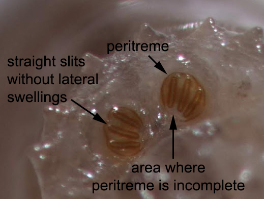
Although we can't see the tracheal trunks to say if they are pigmented or not, the posterior spiracular plate is clearly visualized and shows an incomplete peritreme, relatively-straight slits without lateral swellings, and slits are directed towards the region where the peritreme is incomplete, consistent with Phormia sp.
Some of you mentioned Lucilia sp. in the differential, although in my interpretation, Lucilia has a complete peritreme with distinct button, unlike what we are seeing here.
The CDC has freely-available pictorial keys online which are a useful starting place when examining fly larvae. Blaine and I also published a guide for Laboratory Identification of Arthropod Ectoparasites. If you don't have access to manuscript, send me an email and I can send you a copy for your own personal use.
As Blaine mentioned, this is a case of facultative myiasis involving an existing wound. The wound was caused by removal of the eye due to a large cancerous growth. What I didn't share is that the growth had been neglected, and the wound may have been as well. Several of you mentioned that this is a third stage (L3) larva, which indicates that the larva was allowed to mature for sometime within the wound.
Although we can't see the tracheal trunks to say if they are pigmented or not, the posterior spiracular plate is clearly visualized and shows an incomplete peritreme, relatively-straight slits without lateral swellings, and slits are directed towards the region where the peritreme is incomplete, consistent with Phormia sp.
Some of you mentioned Lucilia sp. in the differential, although in my interpretation, Lucilia has a complete peritreme with distinct button, unlike what we are seeing here.
The CDC has freely-available pictorial keys online which are a useful starting place when examining fly larvae. Blaine and I also published a guide for Laboratory Identification of Arthropod Ectoparasites. If you don't have access to manuscript, send me an email and I can send you a copy for your own personal use.
Subscribe to:
Posts (Atom)