Tuesday, April 12, 2022

Answer to Case 679

 Answer to Parasite Case of the Week 679: Haemaphysalis sp. 

Congratulations to Blaine Mathison, Khaled Itani, Olugbenga Samuel Babatunde, Souti Prasad Sarkhel, Robyn Nadolny, Marc Couturier, @TickReport, and Alvaro Faccini-Martinez for the correct identification! 

It can be tricky to differentiate Haemaphysalis from from Rhipicephalus, so I created the following pictorial guide to help illustrate the key features. 


In the end, it all comes down to a difference of angles - in Haemaphysalis, the outward facing angle is from the palps, whereas with Rhipicephalus, it's from the basis capituli. Both of these ticks also have festoons and their mouthparts are as long as the basis capituli, so they otherwise have a very similar appearance. 

As Blaine points out, there are several members of this genus that are found in the United States, including the newly-invasive H. longicornis, or Asian longhorned tick. There arent' enough features shown in this case to get to the species level, but if you ever have a specimen in your laboratory, you can use this recently published KEY for assistance. Also, if you'd like to learn more about H. longicornis, the CDC has a brief overview HERE. The USDA also has a great site on H. longicornis that includes a regularly-updated map of the tick's distribution throughout the United States.

Thanks again to Florida Fan for donating this case!

Tuesday, April 5, 2022

Case of the Week 678

This week's fascinating and unusual case was donated by Dr. Vicki Schnadig at University of Texas Medical Branch. The patient is an elderly woman from rural Louisiana who presented with a subcutaneous thigh nodule. Dr. Schnadig's team first performed a fine needle aspirate of the lesion which showed the following laminated spherical structure, but no definitive pathogen:

Following this, the team noted a worm-like object protruding from the aspiration site and pulled it out:

The following are histologic sections of this object:


Diagnosis?


Sunday, April 3, 2022

Answer to Case 678

 Answer to the Parasite Case of the Week 678: Sparganosis - infection with the larval form (sparganum) of Spirometra sp.  Kudos to Dr. Francisco Bravo for getting the correct answer, and for the nice description from Florida Fan.

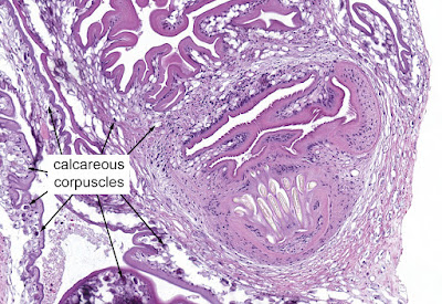
This was a fascinating case as sparganosis is a relatively rare parasitic infection in humans, and the way it was discovered following fine needle aspiration is very unique. It's possible that the laminated calcification seen on the aspirate smear is a calcareous corpuscle, but it's hard to know for sure. Therefore, it was a good thing that the sparganum began to extrude from the aspirate site and could be removed. It's macroscopic and microscopic appearance are diagnostic. Note the longitudinal smooth muscle fibers, loose stroma, and numerous calcareous corpuscles. The latter are seen in all cestodes, in both larval and adult forms. In this case, they mostly appear as empty oval-shaped structures, but can also calcify and appear dark purple on routine H&E stain.

Although removal is curative, this patient should be counseled to avoid eating undercooked fish, reptiles and frogs, to avoid acquiring another case of sparganosis in the future!

Tuesday, March 29, 2022

Case of the Week 677

This week's case came through my lab awhile back and was beautifully captured in the following photographs by Emily Fernholz. The specimen is a Giemsa-stained thin blood film from a patient with travel to Botswana. What parasite is seen here?


Monday, March 28, 2022

Answer to Case 677

Answer to Parasite Case of the Week 677: Plasmodium malariae with classic band and schizont forms. There is also a hint of a basket form, and a nice collection of merozoites that recently burst out of an infected red blood cell. Note that the infected cells are smaller than the uninfected neighboring cells, which is classic for this species. Thank you for all of the great comments!


Tuesday, March 15, 2022

Case of the Week 676

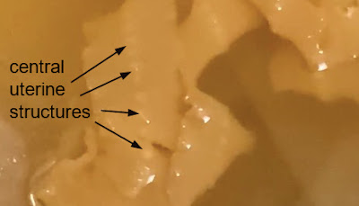
This week's case is generously donated by Dr. Adrienne Showler. The following was passed in stool by an 'adventurous eater', who regularly enjoys raw fish dishes, and also has tried raw beef and pork dishes in the United States. He reports passing similar objects on 4 separate occasions, and the length of each was between 1 and 3 feet. Unfortunately he didn't collect the samples (!), so we only have these photographs:


Likely identification?


Sunday, March 13, 2022

Answer to Case 676

 Answer to the Parasite Case of the Week 676: Cestode, most likely Diphyllobothrium/Dibothriocephalus/Adenocephalus species. The CDC DPDx webpage on diphyllobothriasis explains how some of the names have changed for this group of tapeworms.

Unfortunately the specimen was not submitted to the laboratory and we therefore can't fully examine it. However, central uterine structure seen in the photo below is consistent with an adult tapeworm of the family Diphyllobothriidae:

Thanks again to Dr. Showler for contributing this case!


Sunday, March 6, 2022

Case of the Week 675

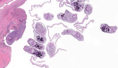
 This week's case was donated by Dr. Paul Valenstein, and it is a beautiful example of an uncommon finding. It is an eyelid biopsy from a 6-year-old African child.


Diagnosis?


Saturday, March 5, 2022

Answer to Case 675

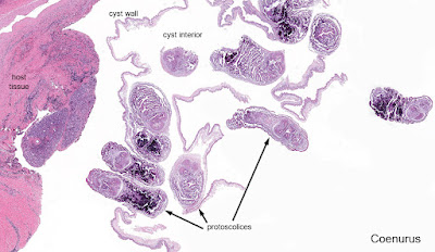
Answer to the Parasite Case of the Week 675: Coenurosis, a tapeworm infection with the larval form of coenurus-forming Taenia species, such as T. multiceps (the most commonly implicated species), T. serialis, T. brauni, and T. glomeratus. 

Some of you noted the resemblance of this tissue infection with cysticercosis. That makes sense since human cysticercosis is due to the related cestode, Taenia solium. The primary difference is the presence of a single protoscolex in cysticercosis, vs. multiple protoscoleces in coenurosis.

Here is my approach to the diagnosis:

  1. We can first tell this is a cestode by the presence of the calcareous corpuscles (nice call Idzi!)
  2. The presence of a thin eosinophilic tegument and loose stroma is also supportive of this being a cestode.
  3. We can further narrow this down to a larval cestode based on its location (within tissue) and cystic appearance. The cystic appearance helps us further narrow down our differential to cystic echinococcosis, cysticercosis, and coenurosis. 
  4. The presence of multiple protoscoleces allows us to rule out cysticercosis (which only has a single protoscolex), bringing us to a differential diagnosis of just 2 cestode infections, cystic echinococcosis and coenurosis. 
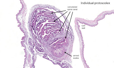
  5. Finally, we can differentiate cystic echinococcus from coenurosis by the appearance of their protoscoleces. Taenia spp. protoscoleces have a convoluted spiral canal, whereas Echinococcus spp. do not.


Tuesday, March 1, 2022

Case of the Week 674

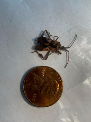
This week's case is courtesy of Dr. Charlie Huskins, one of our pediatric ID physicians. The parents of his patient found the following bug in their home and were concerned that it might be a disease vector. They are concerned that it might have bitten their child, as they have identified a potential 'bite' mark.


Of note, the family lives in the midwestern United States and has no history of recent travel. 

What is the general identification of this bug? Is there any concern for disease transmission?