Monday, October 30, 2017

Answer to Case 466

So many great costumes!
Here are the 'answers' to what they were:

Bobbi and Blaine:
Alexandra:

Jeff and Diana:
Heather and her tick family:

Patty (Lucillia) and Sue (Cochliomyia) botflies:
And a special addition - Mark and Kristine. They weren't at my party (maybe next year?) but deserved to be included here with their awesome costumes:


Monday, October 23, 2017

Case of the Week 465

This week's case is of an elderly woman with thick cutaneous lesions involving the dorsal and ventral aspects of her feet including her toes. There is no associated pain or itching. A sample of the thickened skin was sent to the parasitology laboratory for further examination. Below is what we received; it measured approximately 5 cm in greatest dimension:

Microscopic examination of a small portion of this material showed the following:


Identification? What is the most likely clinical diagnosis?

Sunday, October 22, 2017

Answer to Case 465

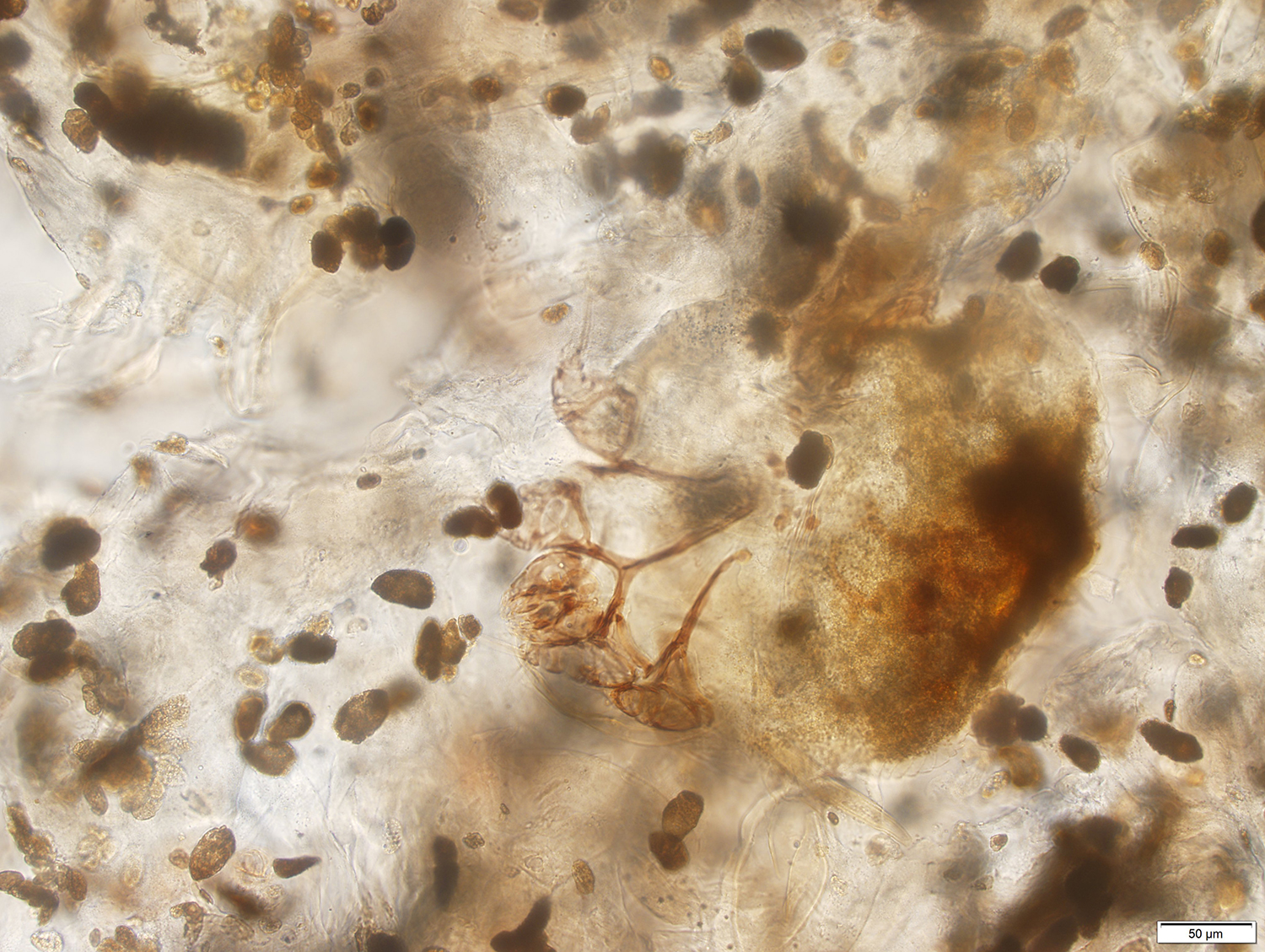
Answer: Sarcoptes scabei infection (scabies); the clinical picture is consistent with crusted ("Norwegian") scabies.

Unlike the classical variant of scabies, crusted scabies does not usually present with severe itching. This is particularly remarkable, given the high mite burden in these cases! The lack of severe itching is related to the depressed immunity of infected individuals. People with crusted scabies are infected with the same mite as people with the classical variant of scabies, but are usually immunocompromised or severely debilitated. Without the host immune response to keep the infection in check, the mites proliferate and produce large crusted lesions that are packed with eggs, mites and fecal pellets.

The predominant finding in this case is the fecal pellets (scybala). Mixed among the scybala are occasional mites which allow for definitive identification:


Monday, October 16, 2017

Case of the Week 464

This week's case was generously donated by Dr. Julie Ribes. The following objects were seen in a Papanicolaou-stained urine specimen from an elderly man with hematuria. They varied in size, measuring ~ 70 micrometers in length. All images were taken using the 40x objective.

Identification?


Sunday, October 15, 2017

Answer to Case 464

Answer: Uric acid crystals

As many of you indicated in your comments, these are NOT Schistosoma haemotobium eggs, despite the superficial resemblance and location in urine, and instead are most consistent with crystals (specifically uric acid crystals). Uric acid crystals can be found in urine in a number of conditions and can be differentiated from S. haematobium eggs using the following features:
  1. Uric acid crystals vary in size and shape and are often much smaller than S. haematobium eggs. In contrast, S. haematobium eggs are regular in size and shape, and quite large (approximately 150 micrometers in length).
  2. Uric acid crystals commonly have points on both ends instead of the single 'pinched-off' spine of S. haematobium eggs. They can also have lateral points or take on other shapes.
  3. There are no internal parasite structures in crystals
  4. Finally, crystals often fracture and break, and may have irregular contours.
I'd encourage you to look at last week's Case 463 to see a good example of S. haematobium eggs. Also, here is a nice side-by-side comparison of a Schistosoma haematobium ovum (left) and a uric acid crystal (right), both stained with Papanicoloau:

I've featured uric acid crystals several times before on this blog, so I thought I would take this opportunity to highlight images from past cases. As you can see from the images below, there is a variety of appearances that uric acid crystals can take in urine:


Tuesday, October 10, 2017

Case of the Week 463

The following objects were seen in a urine specimen obtained from a 16-year old male from Northern Africa. The urine was noted to be grossly bloody. Identification?

They were clearly still alive!


Monday, October 9, 2017

Answer to Case 463

Answer: Schistosoma haematobium ova

The large size (~120 micrometers long), presence of a terminal spine, and location in urine are characteristic of this species, and this identification fits well with the history of hematuria in this patient. The other Schistosoma egg that has a similar appearance is S. intercalatum; in contrast to S. haematobium, it is most commonly found in stool and is somewhat longer (140-170 micrometers). It also has a central bulge, and infection is limited to east central Africa. The patient in this current case is from Northern Africa, outside of the area where S. intercalatum is present.

The video allows you to see the motility of the miracidium inside of the egg, including the "flame cells" (protonephridium). It can be helpful clinically to verify that the eggs are alive, as this indicates an active infection (unless the patient has been recently treated).

 We had a similar case in my lab last year which you can find as Case of the Week 417.

Monday, October 2, 2017

Case of the Week 462

This week I am introducing an exciting new collaboration with Idzi Potters and the Institute of Tropical Medicine Antwerp. This renowned institution has provided health care and research in the field of tropical infectious diseases for more than a century and has accumulated a wealth of marvelous instructive cases. We will share a case from their archives on the first Monday of each Month.

This month's case is of a 60 year-old Belgian woman with a long history of travel to sub-Saharan Africa. She presented with persistent upper abdominal discomfort and radiologic imaging revealed a large liver cyst. Below are representative photographs (shown at 200X to 400X original magnification) and a video clip of the unstained aspirated material. Identification?


See the fascinating motility of one of these objects:


Sunday, October 1, 2017

Answer to Case 462

Answer: Echinococcus sp. protoscoleces, free hooklets and laminated layer. The thick laminated layer is consistent with E. granulosus although the findings would need to be correlated with the clinical and radiologic findings.

This case generated a lot of great comments and discussion! Some of the main features shown in these beautiful photographs by Idzi Potters are the calcareous corpuscles (found in all cestode larvae and adults), protoscoleces with internalized hooklets, free hooklets, and the laminated layer of the cyst wall. The laminated layer is the outer-most layer of the parasite-derived cyst, and is usually surrounded externally by a layer of fibrotic host tissue. Just internal to the laminated layer is the germinal membrane (not easily seen in this case) from which the protoscoleces arise.
The protoscoleces contain an internal ring of hooklets. If they are ingested by a definitive canid host, then the head (scolex) will evert to expose the hooklets; these then aid in attachment to the intestinal lining.
Over time the protoscoleces will degenerate, releasing free hooklets into the cyst fluid. Depending on the state of the cyst, free hooklets may be the only identifiable structures seen. The hard hooklets (both free and within protoscoleces) give a 'gritty' consistency to the aspirated cyst fluid; hence the term "hydatid sand".

Monday, September 25, 2017

Case of the Week 461

This week's case was donated by Dr. Peter Gilligan. The patient is a toddler who presented with high fever and tachycardia. The patient had come to the United States from Uganda 16 months prior to presentation and had not traveled outside of the United States since. The following are representative fields from the peripheral blood smear.


Identification?