Sunday, October 27, 2019

Case of the Week 566

Happy Halloween everyone! In honor of this week, I have photos from my annual Halloween party, and a special 'unknown' from Old One. Feel free to guess what my guests' costumes were:

Me and Felicity with (unplanned) matching costumes. Any ideas what we were?
Me and Old One (so great to meet him in person!)
Heather and her 'life cycle' family
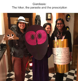
Natalia, Zerelda and Pooja representing the exposure and treatment course for this common parasite:
Another take on this fun parasite by Emily and Nick:
Rebecca, Aimee and Emily demonstrating the woes of being a Clinical Microbiology fellow:
Casey and his wife as another classic parasite:
Kyle and his tapeworm family:
There were many more wonderful costumes that I didn't get pictures of (and many that weren't parasite-related that I will post separately on Facebook). 
And last but not least, HERE is Old One's contribution of a hand-drawn case, animated by his son.


Saturday, October 26, 2019

Answer to Case 566

Answer: lots of fun parasite Halloween costumes; here are the 'answers'


Monday, October 21, 2019

Case of the Week 565

This week's case was generously donated by Professor Agnes Kurniawan from the University of Indonesia. The following motile structure was reported to emerge from the anus of a man from rural Indonesia. He had no other gastrointestinal symptoms.
You can see its subtle movement here:

This structure was expressed from the submitted object:
Identification?

Sunday, October 20, 2019

Answer to Case 565

Answer to Parasite Case of the Week 565: Bertiella species

This case nicely demonstrates a short chain of proglottids and egg (expressed from the proglottids) of Bertiella - a tapeworm of non-human primates that only rarely infects humans. You can read more about this fascinating parasite at the CDC DPDx website.

The proglottids are very short and wide, thus giving this small segment of proglottids the appearance of 'bow tie' pasta as several of my readers pointed out! (arrows point to some of the individual proglottids)
Bertiella eggs have an internal pyriform apparatus which is difficult to make out in this case. It is better seen in my previous case from 2012: Case of the Week 193.
Thanks again to Professor Agnes Kurniawan from the University of Indonesia who donated this case.


Monday, October 14, 2019

Case of the Week 564

This week's case was donated by Blaine Mathison and Marc Couturier. The following forms were seen on peripheral blood smears. No travel history is available at this time. How would you recommend reporting out this case? Are there any additional studies you would recommend?


Sunday, October 13, 2019

Answer to Case 564

Answer: Mixed Plasmodium ovale and Plasmodium falciparum infection.

This was a very nice clear-cut example of a mixed infection. Often they aren't this clear cut! Thanks to everyone who wrote in with the very nice morphologic descriptions supporting the diagnosis.

Santiago noted that "The first five images show many diagnostic morphologic features: the infected RBCs are enlarged, they have an oval shape with jagged edges (fimbriations), Schüffner's dots (fine cytoplasmic stippling) are present in their cytoplasm, and infecting trophozoites are compact; these features are consistent with infection by Plasmodium ovale.

The last two images show banana-shaped gametocytes which are diagnostic of Plasmodium falciparum.

My follow-up question was regarding which additional studies, if any, would be recommended. In this case, the morphology is very convincing, and so diagnostic PCR is likely not needed.  However, it was available in this case, and confirmed the diagnosis of P. falciparum and P. ovale mixed infection. Several of you also correctly noted that quantification of parasitemia is also indicated to guide therapy.

Finally, knowing more about the patient would be important to direct patient care. Information to gather would include where the patient had traveled (to determine if there is circulating resistance to commonly-used antimalarials) and if prior antimalarial therapy had been administered. The fact that only P. falciparum gametocytes were present may indicate that the patient had received prior therapy, since the drugs commonly used to treat P. falciparum are not gametocidal. Thus seeing residual P. falciparum gametocytes is not uncommon after successful treatment.  Now that we know that the patient has P. ovale co-infection, primaquine (or tafenoquine) must be administered to eradicate its hypnotzoite stage (dormant stage in the liver).

Thanks again to Marc and Blaine for donating this case.

Monday, October 7, 2019

Case of the Week 563

It's time for our first case of the month by Idzi Potters and the Institute of Tropical Medicine, Antwerp.   The patient is a 50 yo Belgian patient returning from Italy with intestinal complaints coughs up the following worm:

Identification?

Sunday, October 6, 2019

Answer to Case 563

Answer to Parasite Case of the Week 563: Anisakid larva, not Anisakis species.

As noted by Marc Couturier, this case has a "very nice and clearly defined intestinal caecum [or 'cecum' for my United States readers]. Lips are visible on the worm and the general size would point again to an Anisakidae member. Coughing up and the abdominal pain are helpful clinical correlates as well." Blaine also added some helpful information regarding the diagnostic features of anisakids: "Based on the anteriorly-directed cecum, this is either Pseudoterranova or Contracaecum. Unfortunately it is not possible to definitively tell from the image if a posterior ventricular appendix is present (Contracaecum) or absent (Pseudoterranova). Anisakis species lack both." He also noted that all 3 genera may have an anterior boring tooth.

The arrow in the following image points to the anteriorly-facing cecum.

Thanks again to Idzi for donating this beautiful case. I thought that it nicely complimented last week's case, which had a nice clinical (endoscopy) image but lacked a view of the defining morphologic features.