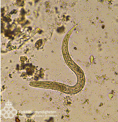
Answer to the Parasite Case of the Week 651: Strongyloides sp. rhabditiform larvae, as evidenced by the short buccal cavity and genital primordium. ALSO in this interesting case are unembryonated and fully embryonated eggs. Eggs are NOT usually shed in the stool in Strongyloides stercoralis infection. So how do we explain these findings? Are these Strongyloides eggs? Or something else? Is there a mixed infection here?
Based on my own interpretation and your comments, I've come up with the 5 possible scenarios to explain the findings in this case:
Scenario 1. Both the larvae and eggs are those of S. stercoralis. As mentioned above, S. stercoralis eggs are not usually shed in stool. However, eggs may rarely be seen in very heavy cases of strongyloidiasis, such as our previous Case of the Week 615 which showed larvae, adults and eggs containing fully embryonated larvae. This scenario would mean that this patient has a potentially serious infection with heavy diarrhea, resulting in passage of eggs before they can hatch in the bowel. As mentioned by Anonymous, it would be helpful to inquire about signs and symptoms of respiratory tract involvement, and if present, examine the sputum for S. stercoralis filariform larvae. Simiarly, Nema suggested obtaining a complete blood count to assess for peripheral eosinophilia, which is commonly seen in cases of hyperinfection.
Scenario 2. We have a MIXED infection, with S. stercoralis larvae (not eggs) AND hookworm eggs. The hookworm egg in the video was fully embryonated, which is unusual for hookworm, but can be seen if the specimen is allowed to sit at room temperature for a while without being placed in fixative.
Scenario 3. A minor variation of the above is that we have a MIXED infection with S. stercoralis eggs (fully embryonated), hookworm eggs (unembryonated), and S. stercoralis rhabditiform larvae.
Scenario 4. A completely different consideration is that this is Strongyloides fuelleborni infection. S. fuelleborni is a zoonotic parasite of non-human primates and humans in Africa and parts of Asia (e.g., Papua New Guinea) in which eggs rather than larvae are shed in stool. The eggs are usually shed in an embryonated state, rather than unembryonated like hookworm, so this doesn't explain why we are also seeing unembryonated eggs. Also, larvae are not usually seen with S. fuelleborni infection. Therefore, this scenario is unlikely.
Scenario 5. Last, but not least, this could be 3-way infection with S. fuelleborni, S. stercoralis, AND hookworm. This unlikely trio would explain the presence of embryonated eggs (S. fuelleborni), unembryonated eggs (hookworm) and Strongyloides sp. rhabditiform larvae (S. stercoralis).
So how do we resolve this conundrum?? Well, we could perform stool PCR or culture to determine the actual identity of the organisms present. However, this is time consuming and these tests are not widely available. Another option is to simply report a probable mixed infection S. stercoralis and hookworm in order to cover all of our bases and ensure that the patient was adequately treated. S. stercoralis is usually treated with ivermectin, whereas hookworm is usually treated with albendazole, mebendazole, or pyrantel pamoate; thus our report could impact the drugs administered.
How would you have handled this case? Feel free to write in and let me know!
UPDATE - we now have PCR confirmation for mixed S. stercoralis and Ancylostoma duodenale!


