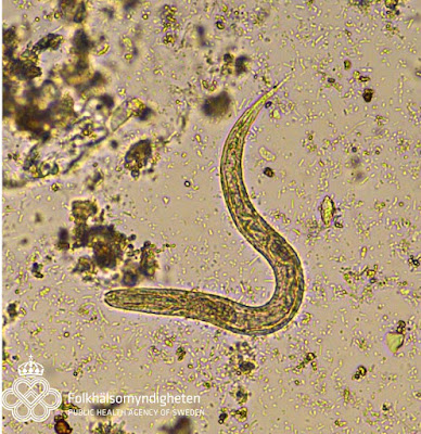
This week's case is from Dr. Ioana Bujila and her colleagues at the Department of Parasitology at the Swedish Public Health Agency. The following were seen in a formalin-ethyl acetate concentration of feces from a young girl with recent travel to India.
Some of these hardy eggs contained live larvae - despite the formalin:
Identification? Any additional tests that you would like to conduct?


9 comments:
Strongyloides stercoralis...thin shell and short buccal cavity....
Nice images! Super cool video!
Thanks for sharing these!
Based on presence of eggs and larvae, differential is between hookworms and Strongyloides.
Depth of buccal cavity is hard to see, but the presence of a genital primordium (2nd picture) makes me lean towards Strongyloides stercoralis.
Size of the eggs is missing, but in view of my ID as strongyloides, they should be around 50 micrometer in length.
The presence of eggs makes be believe that the girl had a heavy diarrhea (short transit time for the eggs).
If it had been hookworm infection, the presence of the larvae would indicate delayed processing of the sample.
Interesting case! Thanks again for sharing!
Looks like Strongyloides stercoralis as egggs are embryonated. Morover, the presence of live larvae in some eggs is in favor of the existence of an endogenous hyperinfectious cycle. It is nécessaire to perform a blood count formula in this young child, in order to assess the proportion of eosinophils. Usually anguilulosis is accompanied by high esoinophilia, while eosinopenia is in favor of malignant anguilulosis,a potentially fatal condition in certain fragile subjects.
Agree with Idzi’s identification. The egg does resemble the ones of hookworm and a stool culture for the filariform larvae would define the identification of Strongyloides stercoralis or hookworm. Filariform larvae of Strongyloides stercoralis have a notched tail, those of hookworm filariforms have a pointed tail.
Florida Fan
If morphology seems compatible with a rhabditoid larva of Strongyloides stercoralis, the presence of eggs containing live larvae make me think to Strongyloides fuelleborni. A DNA sequencing is necessary to confirm it (cox1 I guess?).
Hi everyone,
The eggs where approx. 58x41.
Agree, it is the rhabditiform and egg of Strongyloides stercoralis. Examination of sputum is recommended to assess hyper-infection condition and stool culture for confirmation of Strongyloides and detection of other infection eg. hookworm infection
I would suggest a co-infection of Strongyloides and hookworm. The larvae most certainly look like Strongyloides sp. to me; a couple of the eggs look more like hookworm than Strongy (the one in the video might be a Strongy egg).
I agree with Blaine's co-infection theory. The modulate eggs are within the size range and of similar morphology to hookworms. The larvae are like both hookworm L1, and S. stercoralis L1. If this was a submission to our Veterinary lab, I would culture the specimen for several days and process with a Baerman sedimentation. This would allow any Strongyloides to develop to L3. S. stercoralis L3 can be identified by its long, strait esophagus and double pronged tail, hookworm L3 by having a single tail.
Post a Comment