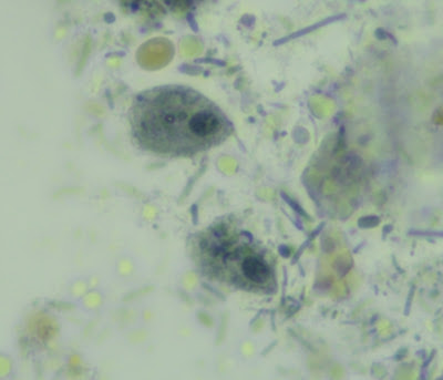
This week's case was generously donated by Dr. Richard Bradbury. The is a permanent mounted stool sample from a Gambian child with watery diarrhea. It is stained with iron haematoxylin; objects of interest are approximately 10-15 micrometers long.
Check out the video for a 3D view and classic motility pattern!


8 comments:
Is it Entamoeba histolytica? I think I see a chromatoid body on the 3D video.
Pentatrichomonas hominis
Here we are, facing another case of Dr. Bradbury. Brace yourself, it typically won’t be a piece of cake. From the video, we can we the very active flagella. This rules out the amoebas whose locomotion is achieved by its pseudopods, the pseudopod projects itself in a direction, pulling with it the very elastic cell wall and then withdrawing with it the entire cell mass. By the size, Pentatrichomonas (formerly Rhetortamonas) hominis is too small to be in the given 10-15 micrometers range. Thus we are left with a possibility: Chilomastix mesnili trophozoites.
As far as the diarrhea is concerned, I believe the condition is due to something else , Chilomastix mesnili is considered a commensal in the intestinal flora, yet its presence indicates a fecal contamination somewhere along the way.
That’s my guess unless Dr. Bradbury intends to give us a lesson on an EID.
Florida Fan
For me, the contenders are Enteromonas hominis, Retortamonas intestinalis, Pentatrichomonas hominis, and Chilomastix mesnili. The first two are ruled out by size and no cysts are evident. I honestly don't think I could tell on trichrome the difference between trophs of Pentatrichomonas and Chilomastix. But we have a beautiful video of undulating movement, rather than rotary movement of Chilo, leading me to Pentatrichomonas. Plus I don't see cysts which further rule out Chilo. So my decision would be Pentatrichomonas hominis. However, if a tech chose any one of these but was wrong due to lack of experience with them, it wouldn't be the end of the world. All are non-pathogens and indicators of ingestion of fecally contaminated food or water.
On a closer look, I can recognize Jebarnes observations. The last video did show not only the flagella but also an undulating membrane running longitudinally. As such, an identification of Pentatrichomonas hominis would be more accurate.
Florida Fan
As two experts told us that the parasite seen on the little video is not pathogenic, but that it indicates the ingestion of fecal contaminated water or food, I can easily believe that this child with a watery diarrhea should be contaminated by anything else, for example by a bacteria (cholera vibrio?) or by a virus (rotavirus?). Anyway this issue should be a public health problem that should be investigated and addressed.
While waiting for you to tell us the end of the story in the event that additional investigations have been undertaken (stool culture), I suggest that the young patient immediately receive ORS, zinc and, if necessary, antibiotics. Additional desirable activities should be carried out, investigating the source of contamination in the environment or surroundings and strengthening WASH interventions in the community where the child comes from.
Perhaps the watery diarrhoea is incidental to the case and is simply flushing trophozoites out from the duodenum, so that they are abundant in the stool and have not have time to degrade before passage? Is there and axostyle visible in any of the slides? These are hints ;)
Post a Comment