Answer: Strongyloides stercoralis
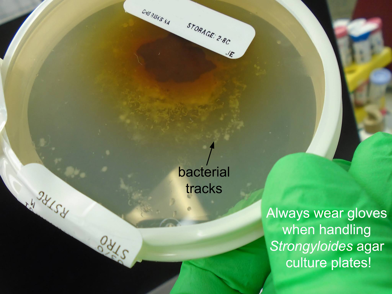
The blood agar and MacConkey plates clearly show bacterial growth tracks that correlate with the migration of larvae through the agar. As noted by Adam, Florida Fan and Arthur, the small size of the larvae, lack of sheath and short buccal cavity are consistent with the rhabditiform larvae of S. stercoralis. Particularly worrying is the fact that the patient is immunosuppressed and thus is at risk of hyperinfection syndrome. Lee commented that the diagnosis is now known and the patient is receiving treatment, so hopefully he will recover.
Thanks again to Lee for sharing this interesting case with us. It's an important reminder to always be on the look-out for larval tracks on bacterial stool cultures. There are a number of organisms that can cause these tracks including hookworm larvae, free-living nematodes and even arthropods (e.g. mites) and it's therefore important to perform a wet prep from the plate to determine the etiology of the tracks.
Showing posts sorted by relevance for query 337. Sort by date Show all posts
Showing posts sorted by relevance for query 337. Sort by date Show all posts
Sunday, February 15, 2015


Sunday, March 15, 2015
Answer to Case 341
Answer: Strongyloidiasis due to Strongyloides stercoralis
As with most biopsies of S. stercoralis, all 3 parasite forms can be seen: the adult female, larvae and eggs:

Note that the eggs resemble those of hookworm. However, unlike human hookworm infection, eggs are never seen in the stool since they hatch to release rhabditiform larvae within the intestine. I like how you can see different stages of cell division within the eggs in this case:

As mentioned by Arthur, this diagnosis fits with the patient's clinical presentation. He states "I would guess that it (the infection) was benign until immunosuppressive chemotherapy began to treat the cancer, at which point a hyperinfective syndrome developed due to the opportunistic and autoinfective nature of this worm. The respiratory failure is likely due to the damage the worms do during the pulmonary stages of the life cycle, and sepsis is well documented in hyperinfective strongyloidiasis due to the filariform larvae penetrating the mucosa to reach the circulatory system, dragging coliform bacteria into the blood stream."
Of note, we take advantage of the propensity for larvae to drag bacteria with them when we use the stool agar culture method to detect Strongyloides stercoralis and other nematodes. When present in a stool specimen, the larvae migrate into the surrounding agar, dragging bacteria with them. The bacteria grow in the tracks, thus producing visible evidence of the larval migration. The agar culture method is the most sensitive means for detecting Strongyloides stercoralis in stool (slightly more sensitive than the older Baermann method - and nicer to perform from a laboratory standpoint because there is no stool in an open container on the benchtop!) You can see some images of S. stercoralis on culture plates in Case of the Week 137, and most recently, by the impressive case donated by Lee (Case of the Week 337). Note that you don't need a special type of agar to see the tracks left by Strongyloides - anything that supports bacterial growth will work. The risk comes from accidental exposure to laboratory techs when they handle the plates with bare hands since Strongyloides stercoralis and hookworm larvae can penetrate intact skin. When we perform the agar culture test in the parasitology lab at my institution, we always tape up the plates and put them in a transparent plastic bag for incubation at room temperature.
As with most biopsies of S. stercoralis, all 3 parasite forms can be seen: the adult female, larvae and eggs:
Note that the eggs resemble those of hookworm. However, unlike human hookworm infection, eggs are never seen in the stool since they hatch to release rhabditiform larvae within the intestine. I like how you can see different stages of cell division within the eggs in this case:
As mentioned by Arthur, this diagnosis fits with the patient's clinical presentation. He states "I would guess that it (the infection) was benign until immunosuppressive chemotherapy began to treat the cancer, at which point a hyperinfective syndrome developed due to the opportunistic and autoinfective nature of this worm. The respiratory failure is likely due to the damage the worms do during the pulmonary stages of the life cycle, and sepsis is well documented in hyperinfective strongyloidiasis due to the filariform larvae penetrating the mucosa to reach the circulatory system, dragging coliform bacteria into the blood stream."
Of note, we take advantage of the propensity for larvae to drag bacteria with them when we use the stool agar culture method to detect Strongyloides stercoralis and other nematodes. When present in a stool specimen, the larvae migrate into the surrounding agar, dragging bacteria with them. The bacteria grow in the tracks, thus producing visible evidence of the larval migration. The agar culture method is the most sensitive means for detecting Strongyloides stercoralis in stool (slightly more sensitive than the older Baermann method - and nicer to perform from a laboratory standpoint because there is no stool in an open container on the benchtop!) You can see some images of S. stercoralis on culture plates in Case of the Week 137, and most recently, by the impressive case donated by Lee (Case of the Week 337). Note that you don't need a special type of agar to see the tracks left by Strongyloides - anything that supports bacterial growth will work. The risk comes from accidental exposure to laboratory techs when they handle the plates with bare hands since Strongyloides stercoralis and hookworm larvae can penetrate intact skin. When we perform the agar culture test in the parasitology lab at my institution, we always tape up the plates and put them in a transparent plastic bag for incubation at room temperature.
Sunday, March 14, 2021
Answer to Case 630
Answer to the Parasite Case of the Week 630: nematode larvae; primary differential is Strongyloides sp., hookworm, Trichostrongylus, and free-living nematodes (e.g., Rhabditis sp.).
The agar plate culture (a.k.a., Koga plate) is a relatively safe and straight-forward method to increase detection of S. stercoralis in feces, and can also be used to culture other nematodes by allowing the eggs in feces to hatch and mature. The procedure is performed by placing a small amount of stool (as shown here) in the center of a nutrient agar. Any agar that supports the growth of enteric bacteria will do, including sheep blood agar and Mueller Hinton agar. We make our own in-house agar using beef broth. If larvae are present in the specimen, they will move over the agar and carry bacteria from the feces with them. The bacteria grow in their tracks, leaving a visible trail of their journeys. Our technologists examine the plates daily for these serpiginous tracks of bacteria. If they are seen, they examine the plate under a light microscope (4x or 10x objective) to look for larvae. HERE is a link to the publication by Dr. Koga detailing the modified agar culture method that we use in our lab.
This case didn't have very pronounced bacterial tracks. However, you can see great examples in some of my previous cases:
Case of the Week 110 and Answer (S. stercoralis in sputum; microscopic track on our in-house agar)
Case of the Week 112 and Answer (Hookworm in a duodenal aspirate; very clear track on our in-house agar)
Case of the Week 137 and Answer (S. stercoralis tracks on sheep blood agar; this was an incidental finding on bacterial stool culture and shell vial viral culture)
Case of the Week 151 and Answer (video of S. stercoralis larvae and tracks on our in-house agar)
Case of the Week 218 and Answer (video of S. stercoralis larvae in agar)
Case of the Week 337 and Answer (S. stercoralis; incidental finding on bacterial stool culture: sheep blood agar and McConkey agar)
Wow, I didn't realize how many cases I've posted of this over the years!
As Marc mentions, the next step is to examine the larvae to identify the genus present, since other nematode eggs in the stool can hatch over a period of days and produce similar-appearing larvae. Free-living larvae may also be present if the fecal specimen was contaminated with soil (which would be unlikely in this case).
To safely examine the larvae, we flood the plate with formalin and let it sit for several minutes. We then carefully remove some of the peripheral material on the plate and place it on a slide to examine microscopically.
This case was particularly remarkable because many of the larvae had matured to be free-living Strongyloides adults. We saw both males and gravid females! You can actually see them on the agar itself, which is really impressive. We suspect that the stool was not submitted immediately after passage by the patient, thus explaining the presence of these advanced forms.
Gravid free-living females:


Male

Adults seen on the plate:

The last question to answer is whether these are S. stercoralis or S. fuelleborni - but that is a subject for a future post!
Monday, February 16, 2015
Case of the Week 337
The following case was generously donated by Lee. The patient is a 26 year old male with advanced testicular cancer who developed abdominal pain after several rounds of immunosuppressive chemotherapy. A stool specimen submitted for bacterial enteric pathogens culture produced the following pattern on blood agar plate (BAP) and MacConkey (MAC) agar:
BAP

MAC agar

Wet preps from the plate:


BAP
MAC agar
Wet preps from the plate:
Subscribe to:
Posts (Atom)


