
Monday, December 30, 2019
Sunday, December 29, 2019
Answer to Case 575
Answer: Ctenocephalides canis. This nice little male flea has characteristic pronotal and genal combs, and a more rounded head than C. felis. There are other important differentiating characteristics as well (you can read about them HERE) and so identification to the species level is best performed by experts.
Thanks to Anon for the lovely poem. I should have thought of this for Christmas!
Anonymous said...
a little late, I would have preferred a fleas navidad..
fleas navidad, fleas navidad
oy contraro fleas are so bad.
I want to wish you a itchy Christmas
I want to wish you a itchy Christmas
from the bottom of my scratch...
Happy New year, all....
Wishing you all a very happy, healthy and productive New Year, full of fascinating parasite cases!
Bobbi (a.k.a. ParasiteGal)
Monday, December 23, 2019
Case of the Week 574
Happy Holidays to all of my readers! Can anyone tell me who this little arthropod is?


Sunday, December 22, 2019
Answer to Case 574
Answer to Parasite Case of the Week 574: Holiday bedbug, most likely Cimex lecturlaris. We seem to have some 'controversy' about the nomenclature here; Eagleville identified this as Cimex lectularius (subspecies Santaclausus), whereas Idzi identified it as Cimex santaclarius. I will rely on my taxonomy experts to tell us which is correct 😉.
For now, I'll leave you with this festive poem from Blaine:
For now, I'll leave you with this festive poem from Blaine:
Dashing through the sheets
on six segmented feet
O'er the pillows they go
sucking blood all the way
lap lap lap
Pointed beak goes down
after you turn off the lights
oh what fun it is to suck
blood from a host tonight
Oh, bed bugs suck
bed bugs suck
bed bugs suck your blood
Just be glad
they don't spread disease
or any nasty crud!
Monday, December 16, 2019
Case of the Week 573
This week's case is Dr. Charles (Chuck) Sturgis. He noted the following structures on a Papanicolaou-stained anal Pap smear (performed for cancer screening). They measure approximately 14 micrometers in length. Identification?


Sunday, December 15, 2019
Answer to Case 573
Answer to the Parasite Case of the Week 573: Giardia duodenalis (a.k.a. G. lamblia, G. intestinalis) cysts
This is the third time I've seen this finding in an anal pap smear - each time it was an unexpected, incidental finding. Despite this being a stain not commonly used in the parasitology laboratory, all of the key morphologic features including the nuclei, central axonemes, and curved median bodies, can be seen.

This is the third time I've seen this finding in an anal pap smear - each time it was an unexpected, incidental finding. Despite this being a stain not commonly used in the parasitology laboratory, all of the key morphologic features including the nuclei, central axonemes, and curved median bodies, can be seen.

Monday, December 9, 2019
Case of the Week 572
This week's cool case is from Dr. Marta Maia. The specimen is skin currettings, and the object below was viewed using a dissecting microscope. The patient had developed a firm, painful 3 mm-diameter lesion on the sole of his foot after a recent vacation in Brazil. During his vacation, we swam in the ocean and walked barefoot on the beach. Identification?


Sunday, December 8, 2019
Answer to Case 572
Answer to Parasite Case of the Week 572: Tungiasis, due to the parasitic female sand flea. As Blaine mentioned, it is most likely Tunga penetrans, but in Brazil, Ecuador and Peru, there is a second species infesting humans: T. trimamillata. It's not possible to tell the two species apart from this image alone.
What we can see from this beautiful image by Dr. Maia is an anterior portion of the female Tunga flea that had been curretted from the patient's foot lesion.

Infestation with the Tunga flea is called tungiasis. From anonymous: Tungiasis "is one of our favorite words as sounds very nasty and rolls off the tongue very nicely." Indeed, tungiasis is a particularly nasty, painful, and potentially debilitating condition where the female flea burrows head first into exposed skin - often between the toes and under nail beds. Santiago reminds us that medical treatment is ineffective, and "prevention is thus the best way of controlling the disease, preventing the parasite from penetrating the intact skin. But who wants to wear shoes at the beach?"
Thanks again to Dr. Maia for sharing this case!
Monday, December 2, 2019
Case of the Week 571
Welcome to the first case of the month, a regular feature by Idzi Potters and the Institute of Tropical Medicine, Antwerp. The following objects were seen in an unstained duodenal aspirate specimen. Identification?


Sunday, December 1, 2019
Answer to Case 571
Answer: Giardia duodenalis (a.k.a. G. lamblia, G. intestinalis) trophozoites with "falling leaf" or "tumbling" motility. Note that this is quite different than the "spiraling" motility of Chilomastix mesnili and the "jerky" motility of Pentatrichomonas hominis - two other flagellates - both non-pathogns - that may be seen in human stool specimens. As pointed out by Florida Fan, the motility is further enhanced by dark field videography, which makes everything more interesting. Thanks again to Idzi for the very cool videos!
Santiago gave us further information on how Giardia trophozoites move in the intestine to attach to the duodenal intestinal mucosa:
After excystation in the small intestine, the trophozoites quickly swim towards the epithelium and attach forming a monolayer; this contributes to the pathology and allows the parasite to escape the turbulent flow of the small intestine and continue the life cycle in the human host.
To achieve this, it uses a combination of movements involving its four pairs of flagella as well as its caudal region, and it is able to switch its motility from "free swimming" in the intestinal lumen, which is more rapid, to a "pre-attachment" pace which is slower and more stable, facilitating effective attachment to the intestinal epithelium in the desired location to form a monolayer.
Lastly, Old One reminds us how
Antonie van Leeuwenhoek was delighted when he took a look
at his own watery stool
Seeing For the very first time
Animalcules
“Bellies flatlike with bodies furnisht with sundry little paws. making quick motion with these paws, yet for all that, they made but slow progress yet a-moving very prettily.”
What a neat parasite!
Monday, November 25, 2019
Case of the Week 570
This week's case was generously donated by Dave Neitzel at the Minnesota Department of Health. A mutant headless tick! Wouldn't it be great if they were all this way?

 Can anyone identify the genus of this tick?


Sunday, November 24, 2019
Answer to Case 570
Answer to Parasite Case of the Week 570: Ixodes scapularis adult female with a missing external capitulum and mouthparts. Or as Blaine noted, "A headless tick: Ichabod scapularis!"
The image below shows these parts where they are normally located on the accompanying female tick (white oval):
 Examination of the ventral aspect shows an "inverted-U" shaped anal groove, allowing this tick to be identified to the Ixodes genus. One reader had queried if the genital pore was also absent on our mutant tick, but re-examination by Dave Neitzel indicated that it IS there; it is just obscured by the glare of the light.
So how did this happen? Well, Dave and some of our readers rightly indicated that the tick must have taken 2 prior blood meals in order to molt from larva to nymph, and then nymph to adult; thus it must have had functional mouthparts during those previous stages. However, as Dave noted, "something went horribly wrong during the last mold from nymph to adult. Given the radical changes that occur during a molt, I am surprised that we don't see more obvious mutations!" He also shared that he has seen several 7-legged ticks from his regular field collections in Minnesota, but surprisingly, no other mutants. He hypothesized that the capitulum and mouthparts are still present, but that they are inverted and therefore not externally-facing. The lack of of the visible capitulum and mouthparts has shifted the position of the legs, thus making them look longer. Finally, he commented on the "sclerotic structure" noted between the first pair of the legs by Makrone. He also had seen this and speculated that it might be some of the residual tissue from the inverted mouthparts/capitulum.
Thanks to Dave for the fascinating case! As Sam mentioned, Ixodes scapularis is the vector for Babesia microti, as well as several bacterial pathogens and Powassan virus. Wouldn't it be great if they were all 'headless'?
The image below shows these parts where they are normally located on the accompanying female tick (white oval):

So how did this happen? Well, Dave and some of our readers rightly indicated that the tick must have taken 2 prior blood meals in order to molt from larva to nymph, and then nymph to adult; thus it must have had functional mouthparts during those previous stages. However, as Dave noted, "something went horribly wrong during the last mold from nymph to adult. Given the radical changes that occur during a molt, I am surprised that we don't see more obvious mutations!" He also shared that he has seen several 7-legged ticks from his regular field collections in Minnesota, but surprisingly, no other mutants. He hypothesized that the capitulum and mouthparts are still present, but that they are inverted and therefore not externally-facing. The lack of of the visible capitulum and mouthparts has shifted the position of the legs, thus making them look longer. Finally, he commented on the "sclerotic structure" noted between the first pair of the legs by Makrone. He also had seen this and speculated that it might be some of the residual tissue from the inverted mouthparts/capitulum.
Thanks to Dave for the fascinating case! As Sam mentioned, Ixodes scapularis is the vector for Babesia microti, as well as several bacterial pathogens and Powassan virus. Wouldn't it be great if they were all 'headless'?
Monday, November 18, 2019
Case of the Week 569
This week's case was captured by my awesome Parasitology Education Specialist, Felicity Norrie, MLS(ASCP). The following were identified from skin scrapings from a resident of a skilled nursing facility. Identification?


Sunday, November 17, 2019
Answer to Case 569
Answer to Parasite Case of the Week 569: Sarcoptes scabei eggs, larva and adult. Fecal pellets (scybala) are also seen. I love how you can see the larval mite emerging from the egg in this photo:

As several readers mention, scabies is infectious to others, and thus the residents and health care workers in this skilled nursing facility must be evaluated for infection and treated if need be. Anonymous also mentioned that "scabies is not a problem of lack of hygiene but of overcrowding and wherever close person-to person contact is common. Scabies spreads quickly especially in nursing/care homes if no skilled GP or dermatologist is available to diagnose the index Patient, even in highly developed countries." This comments brings up the important point that infection does NOT reflect poor hygiene of the infected individuals. The mite infects all ages, ethnicities and social strata.

As several readers mention, scabies is infectious to others, and thus the residents and health care workers in this skilled nursing facility must be evaluated for infection and treated if need be. Anonymous also mentioned that "scabies is not a problem of lack of hygiene but of overcrowding and wherever close person-to person contact is common. Scabies spreads quickly especially in nursing/care homes if no skilled GP or dermatologist is available to diagnose the index Patient, even in highly developed countries." This comments brings up the important point that infection does NOT reflect poor hygiene of the infected individuals. The mite infects all ages, ethnicities and social strata.
Monday, November 11, 2019
Case of the Week 568
This week's case is from Old One - illustrated by him and animated by his son. It features an arthropod that measures a few millimeters in length.
 See it in action HERE!
Identification?

Identification?
Sunday, November 10, 2019
Answer to Case 568
Answer to Parasite Case of the Week 568: Pseudoscorpion or false scorporion, not a human parasite.
This fun little arthropod is occasionally submitted to the clinical laboratory for identification, and may be mistaken as a true scorpion. While both scorpions and pseudoscorpions are arachnids, pseudoscorpions are very small (1 cm or less in length) and lack a tail with a stinger. As sylvie g and Santiago note, pseudoscorpions can occasionally be found in the house, but they don't bite or sting humans, and instead feed on other small arthropods such as booklice.

Thanks again to Old One and his son for the donation of the cool animated illustration.
Tuesday, November 5, 2019
Case of the Week 567
This week's case is from Idzi Potters and the Institute of Tropical Medicine, Antwerp. The following object was seen in a concentrated stool specimen from a 3-year-old toddler with diarrhea (40x objective). It measures 45 micrometers in greatest dimension. Identification?


Monday, November 4, 2019
Answer to Case 567
Answer: Hymenolepis nana egg.
As many of you pointed out, this eggs beautifully demonstrates the filaments arising from the 2 poles of the inner membrane (arrow heads, below image). You can't make it out here, but there are 4-8 of these filaments arising from each pole. You can also nicely see the hooks of the 6-hooked oncosphere:
 Some of you may know that humans become infected with this parasite when they ingest infected arthropods. As Sam mentioned, children are a common host - likely due to their tendency to put things in their mouth. Adults can also become infected as Blaine reminds us in his poem:
If you do an internet search for "oatmeal" and "bugs," you will find multiple consumer complaints (and some nice YouTube videos) to explain the cereal reference.
Importantly, infection via ingestion of eggs shed in stool (autoinfection or from another person) is also possible, making this a common intestinal cestode infection worldwide.
As many of you pointed out, this eggs beautifully demonstrates the filaments arising from the 2 poles of the inner membrane (arrow heads, below image). You can't make it out here, but there are 4-8 of these filaments arising from each pole. You can also nicely see the hooks of the 6-hooked oncosphere:

There once was a chap from Indiana
who with his cereal enjoyed a banana
but with the cereal he did eat
he received an unexpected treat
The cestode known as Hymenolepis nana!
If you do an internet search for "oatmeal" and "bugs," you will find multiple consumer complaints (and some nice YouTube videos) to explain the cereal reference.
Importantly, infection via ingestion of eggs shed in stool (autoinfection or from another person) is also possible, making this a common intestinal cestode infection worldwide.
Sunday, October 27, 2019
Case of the Week 566
Happy Halloween everyone! In honor of this week, I have photos from my annual Halloween party, and a special 'unknown' from Old One. Feel free to guess what my guests' costumes were:
Me and Felicity with (unplanned) matching costumes. Any ideas what we were?


Me and Felicity with (unplanned) matching costumes. Any ideas what we were?

Me and Old One (so great to meet him in person!)

Heather and her 'life cycle' family

Natalia, Zerelda and Pooja representing the exposure and treatment course for this common parasite:

Another take on this fun parasite by Emily and Nick:

Rebecca, Aimee and Emily demonstrating the woes of being a Clinical Microbiology fellow:

Casey and his wife as another classic parasite:

Kyle and his tapeworm family:

There were many more wonderful costumes that I didn't get pictures of (and many that weren't parasite-related that I will post separately on Facebook).
And last but not least, HERE is Old One's contribution of a hand-drawn case, animated by his son.
Saturday, October 26, 2019


Monday, October 21, 2019
Case of the Week 565
This week's case was generously donated by Professor Agnes Kurniawan from the University of Indonesia. The following motile structure was reported to emerge from the anus of a man from rural Indonesia. He had no other gastrointestinal symptoms.

This structure was expressed from the submitted object:
 Identification?

You can see its subtle movement here:
This structure was expressed from the submitted object:

Sunday, October 20, 2019
Answer to Case 565
Answer to Parasite Case of the Week 565: Bertiella species
This case nicely demonstrates a short chain of proglottids and egg (expressed from the proglottids) of Bertiella - a tapeworm of non-human primates that only rarely infects humans. You can read more about this fascinating parasite at the CDC DPDx website.
The proglottids are very short and wide, thus giving this small segment of proglottids the appearance of 'bow tie' pasta as several of my readers pointed out! (arrows point to some of the individual proglottids)
 Bertiella eggs have an internal pyriform apparatus which is difficult to make out in this case. It is better seen in my previous case from 2012: Case of the Week 193.
 Thanks again to Professor Agnes Kurniawan from the University of Indonesia who donated this case.
This case nicely demonstrates a short chain of proglottids and egg (expressed from the proglottids) of Bertiella - a tapeworm of non-human primates that only rarely infects humans. You can read more about this fascinating parasite at the CDC DPDx website.
The proglottids are very short and wide, thus giving this small segment of proglottids the appearance of 'bow tie' pasta as several of my readers pointed out! (arrows point to some of the individual proglottids)


Monday, October 14, 2019
Case of the Week 564
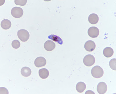
This week's case was donated by Blaine Mathison and Marc Couturier. The following forms were seen on peripheral blood smears. No travel history is available at this time. How would you recommend reporting out this case? Are there any additional studies you would recommend?


Sunday, October 13, 2019
Answer to Case 564
Answer: Mixed Plasmodium ovale and Plasmodium falciparum infection.
This was a very nice clear-cut example of a mixed infection. Often they aren't this clear cut! Thanks to everyone who wrote in with the very nice morphologic descriptions supporting the diagnosis.
Santiago noted that "The first five images show many diagnostic morphologic features: the infected RBCs are enlarged, they have an oval shape with jagged edges (fimbriations), Schüffner's dots (fine cytoplasmic stippling) are present in their cytoplasm, and infecting trophozoites are compact; these features are consistent with infection by Plasmodium ovale.

The last two images show banana-shaped gametocytes which are diagnostic of Plasmodium falciparum.
My follow-up question was regarding which additional studies, if any, would be recommended. In this case, the morphology is very convincing, and so diagnostic PCR is likely not needed. However, it was available in this case, and confirmed the diagnosis of P. falciparum and P. ovale mixed infection. Several of you also correctly noted that quantification of parasitemia is also indicated to guide therapy.
Finally, knowing more about the patient would be important to direct patient care. Information to gather would include where the patient had traveled (to determine if there is circulating resistance to commonly-used antimalarials) and if prior antimalarial therapy had been administered. The fact that only P. falciparum gametocytes were present may indicate that the patient had received prior therapy, since the drugs commonly used to treat P. falciparum are not gametocidal. Thus seeing residual P. falciparum gametocytes is not uncommon after successful treatment. Now that we know that the patient has P. ovale co-infection, primaquine (or tafenoquine) must be administered to eradicate its hypnotzoite stage (dormant stage in the liver).
Thanks again to Marc and Blaine for donating this case.
Monday, October 7, 2019
Case of the Week 563
It's time for our first case of the month by Idzi Potters and the Institute of Tropical Medicine, Antwerp. The patient is a 50 yo Belgian patient returning from Italy with intestinal complaints coughs up the following worm:


Identification?
Sunday, October 6, 2019
Answer to Case 563
Answer to Parasite Case of the Week 563: Anisakid larva, not Anisakis species.
As noted by Marc Couturier, this case has a "very nice and clearly defined intestinal caecum [or 'cecum' for my United States readers]. Lips are visible on the worm and the general size would point again to an Anisakidae member. Coughing up and the abdominal pain are helpful clinical correlates as well." Blaine also added some helpful information regarding the diagnostic features of anisakids: "Based on the anteriorly-directed cecum, this is either Pseudoterranova or Contracaecum. Unfortunately it is not possible to definitively tell from the image if a posterior ventricular appendix is present (Contracaecum) or absent (Pseudoterranova). Anisakis species lack both." He also noted that all 3 genera may have an anterior boring tooth.
The arrow in the following image points to the anteriorly-facing cecum.

Thanks again to Idzi for donating this beautiful case. I thought that it nicely complimented last week's case, which had a nice clinical (endoscopy) image but lacked a view of the defining morphologic features.
As noted by Marc Couturier, this case has a "very nice and clearly defined intestinal caecum [or 'cecum' for my United States readers]. Lips are visible on the worm and the general size would point again to an Anisakidae member. Coughing up and the abdominal pain are helpful clinical correlates as well." Blaine also added some helpful information regarding the diagnostic features of anisakids: "Based on the anteriorly-directed cecum, this is either Pseudoterranova or Contracaecum. Unfortunately it is not possible to definitively tell from the image if a posterior ventricular appendix is present (Contracaecum) or absent (Pseudoterranova). Anisakis species lack both." He also noted that all 3 genera may have an anterior boring tooth.
The arrow in the following image points to the anteriorly-facing cecum.
Thanks again to Idzi for donating this beautiful case. I thought that it nicely complimented last week's case, which had a nice clinical (endoscopy) image but lacked a view of the defining morphologic features.
Monday, September 30, 2019
Case of the Week 562
This week's case was generously donated by Dr. David Hernandez Gonzalo. The patient is a middle-aged male who presented with abdominal discomfort and nausea. An endoscopy was performed which revealed the following object in the gastric antrum:
 The worm was sent to pathology and was sectioned, revealing the following (H&E):

 Identification?


Subscribe to:
Posts (Atom)


